
Overview
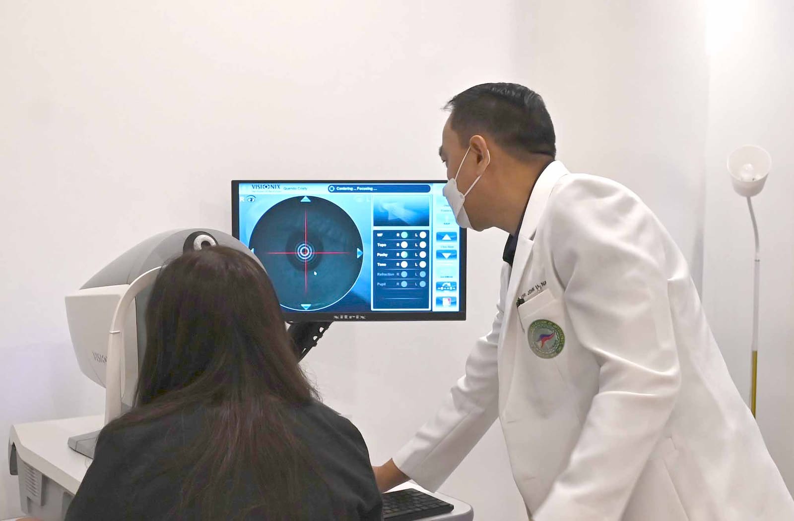
Corneal topography is an advanced diagnostic imaging technique that creates a detailed three-dimensional map of your cornea's surface curvature. Like a topographical map shows the contours of land, corneal topography reveals the hills, valleys, and irregularities of your cornea—the clear front surface of your eye.
The cornea is responsible for about 70% of the eye's focusing power, so even small irregularities can significantly affect vision quality. Topography can detect conditions like keratoconus, corneal warpage from contact lenses, and subtle irregularities that may not be apparent during standard examination.
This technology is essential for fitting specialty contact lenses, planning refractive surgery, and monitoring corneal conditions over time. The detailed maps produced help us understand exactly how your cornea is shaped and how it affects your vision, enabling more precise and personalized treatment approaches.
What's Included
Comprehensive features and services included in this examination.
Who Should Consider This Service?
This service is ideal for the following individuals.
Age Group
- Teenagers (keratoconus screening)
- Young adults considering LASIK
- Adults with irregular astigmatism
- Contact lens wearers
Symptom-Based
- Distorted or ghosted vision
- Frequent prescription changes
- Poor vision despite glasses
- Contact lens discomfort
Medical-Based
- Suspected keratoconus
- Post-corneal surgery monitoring
- Corneal dystrophies
- Irregular astigmatism
Lifestyle-Based
- LASIK/PRK candidates
- Specialty contact lens needs
- Ortho-K candidates
- Those with hard-to-fit eyes
What to Expect
A step-by-step guide to your appointment experience.
Preparation
Contact lenses should be removed before testing (soft lenses 3+ days, rigid lenses 2+ weeks for most accurate results). No drops are needed.
Positioning
You'll sit comfortably with your chin and forehead resting on supports, looking at a target in the center of the instrument.
Image Capture
The topographer projects rings of light onto your cornea and captures how they reflect. This takes just seconds and involves no contact with your eye.
Map Generation
The computer analyzes the reflected patterns and generates detailed color-coded maps showing corneal curvature across thousands of points.
Analysis
The doctor reviews the maps, looking for patterns that indicate normal corneas, astigmatism, keratoconus, or other conditions.
Results Discussion
We explain what the maps show about your cornea and how this information guides your treatment or lens fitting.
Technology & Equipment
State-of-the-art equipment used for accurate diagnosis and treatment.
Placido Disc Topographer
Projects concentric rings onto the cornea and analyzes their reflection to map surface curvature with high precision.
Scheimpflug Imaging
Advanced technology that images the cornea from multiple angles, providing both front and back surface measurements.
Elevation Mapping
Shows how the corneal surface deviates from a perfect sphere, essential for detecting subtle abnormalities.
Keratoconus Detection Software
Specialized algorithms that analyze topography patterns to identify early keratoconus before it becomes clinically obvious.
Frequently Asked Questions
Common questions about this service answered.
Why Choose Nakpil Eye Clinic?
Experienced Team
15+ years of expertise in eye care
Advanced Technology
State-of-the-art diagnostic equipment
Personalized Care
Tailored treatment plans for each patient
Comprehensive Services
Full range of eye care solutions